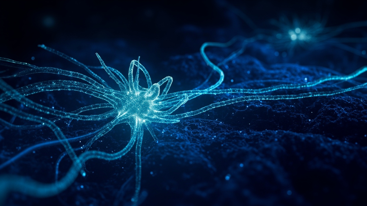
TL;DR: In mice, switching on rare cortical Sst-Chodl neurons was sufficient to synchronize cortex-wide activity and promote sleep.
A recent Nature study from neuroscientists Geoffrey Terral and Renata Batista-Brito at the Albert Einstein College of Medicine in New York identifies a tiny cortical population that can drive the brain toward sleep, according to Ars Technica. The cells, termed Sst-Chodl neurons, represent only around one percent of inhibitory neurons in the cortex, yet direct activation in mice pushed network activity into sleep-like coordination and the animals fell asleep. The result reframes the cerebral cortex from a passive recipient of deep-brain sleep commands into a structure capable of initiating sleep rhythms on its own.
The work was reported on Sept. 18, 2026 by Ars Technica, with independent coverage of the same mouse experiments published Sept. 15, 2026 by Medical Daily and Sept. 20, 2026 by Live Well Editorial Team. All of the manipulation data come from mice. According to Medical Daily, similar cells are present across mammals including humans, but that conservation does not demonstrate an identical sleep-promoting role in people.
A different account of where sleep starts
For a long time, sleep research treated the cerebral cortex as a follower that reacted to signals from deep structures, according to Ars Technica. In that conventional view, subcortical regions controlled sleep and arousal, while the cortex displayed the slow rhythms of deep sleep without generating the trigger.
Terral told Ars Technica that sleep is usually associated with control by subcortical regions. The new experiments challenge that division by showing that activation of a cortical cell class alone is sufficient to promote sleep in freely moving mice. Terral said the findings show the cortex can initiate the rhythm by itself, not merely display it.
Medical Daily described the same shift, noting that sleep has often been studied as a state governed largely by deeper arousal-regulating structures that then influence the cortex. The new results, it reported, indicate the cortex has a more active part in organizing the change from wakefulness to sleep.
The rare cells at the center
The neurons are defined by activity of two genes, somatostatin and chondrolectin, according to the Live Well Editorial Team. The label Sst-Chodl reflects that dual genetic signature. They belong to the inhibitory class, meaning their output quiets other cells rather than exciting them.
They are extremely sparse. Ars Technica reported that they constitute about one percent of cortical inhibitory neurons. Because inhibitory neurons account for about one-fifth of all cortical neurons, the Sst-Chodl group amounts to roughly one neuron in a thousand in cortex. Medical Daily placed the share at about 0.2% of cells in the cerebral cortex.
That scarcity created a technical barrier. Targeting either gene alone captures a large and mixed family of neurons, according to Ars Technica. Batista-Brito said a single gene is not able to target these cells, and warned that without precise selection, unrelated cells would dominate the sample. Her group spent years constructing a labeling approach that marks a cell only when both genes are active.
Batista-Brito described the effort as high-risk because the chance of detecting a signal from 0.1 percent of cortical neurons appeared very low, according to Ars Technica. She said multiple grant proposals on the project were rejected as too high-risk.
Wiring built for broadcast
The first anatomical views justified the effort. Batista-Brito told Ars Technica that she initially saw only two or three cell bodies in the whole brain, yet observed extensive branching throughout the visual cortex unlike anything she had seen in another neuron.
That branching, described as arborization, forms tree-like extensions that link a neuron into wider communication networks. The pattern was striking because inhibitory neurons are almost always local, according to Ars Technica. A typical cortical inhibitory cell receives inputs from different and sometimes distant regions but controls only its immediate neighborhood.
Batista-Brito said Sst-Chodl neurons reverse that arrangement, receiving relatively precise inputs while broadcasting information widely. According to Ars Technica, a single Sst-Chodl cell can ramify through the entire visual cortex and send axons toward areas involved in touch, hearing, spatial memory, navigation and voluntary movement.
Medical Daily emphasized the same contrast, reporting that unlike many neurons that communicate mainly nearby, these cells maintain long connections across cortex. That reach, it reported, gives a very small population the potential to influence activity over a much larger territory. The Live Well Editorial Team likewise noted that most neocortical inhibitory neurons act locally, while these cells exert widespread influence through long-range axons that contact multiple regions simultaneously.
Active during drowsiness, quiet during alert movement
To determine when the cells are naturally engaged, the team imaged them while simultaneously monitoring pupil size, muscle tone, facial movement, running and cortical electrical activity, according to Ars Technica. Of 111 imaged cells, 95 became active during slow-wave sleep and quiet, motionless wakefulness, and became silent during running and REM sleep.
Medical Daily reported a compatible pattern, stating that the neurons were relatively quiet while animals were awake, increased activity as mice became drowsy and entered non-REM sleep, and that cortical electrical activity simultaneously slowed and became more synchronized.
During slow-wave sleep, cortical networks alternate between UP states of vigorous firing and DOWN states of near-silence. Terral said Sst-Chodl activity rose even further as an UP state ended, increasing while other neurons decreased, according to Ars Technica. He also said the cells skip the rebound displayed by surrounding cells when networks exit a DOWN state, behaving differently from every other measured cell around that transition.
The Live Well Editorial Team summarized the selectivity in behavioral terms, reporting that the cells were selectively active during low-arousal states and mostly silent during high arousal.
Turning the cells on produces sleep rhythms
Observation alone could not show causation, so the researchers used optogenetics, a method that introduces a gene for a light-activated ion channel to make selected nerve cells fire on command, according to Ars Technica. Medical Daily said the team then tested the causal question by activating Sst-Chodl cells directly.
Stimulation delivered in visual cortex increased delta power, defined as the slowest, highest-amplitude waves measured during deep restorative sleep, across every cortical layer, according to Ars Technica. It also tightened spike timing and made DOWN states more frequent and longer, while overall firing changed little.
Terral said the neurons alter coordination more than firing rate, determining whether neurons fire together rather than how often they fire, according to Ars Technica. The induced oscillation did not follow the drive frequency. Batista-Brito said flat stimulation, delta-frequency stimulation, 20-hertz stimulation and 60-hertz stimulation all produced the same kind of oscillation, pointing to intrinsic cellular properties that favor one frequency. She added that the mechanism behind that preference is not currently known.
The spatial effect extended well beyond the stimulation site. Terral said cell bodies located in visual cortex could regulate most recorded neurons, according to Ars Technica. Direct inhibitory currents appeared in one-third of cells two millimeters away, and stimulation in visual cortex produced measurable effects as distant as frontal motor areas.
Mice fell asleep more readily
In behavioral trials with freely moving animals, widespread activation increased slow-wave and REM sleep, according to Ars Technica. Medical Daily reported that stimulated mice fell asleep more readily and spent more time asleep, with neocortical activity shifting toward slower synchronized patterns normally linked to sleep.
The Live Well Editorial Team described selective activation as sufficient to promote multi-region cortical synchronization characteristic of low-arousal states and to induce sleep. That distinction matters because it separates cells that merely accompany sleep from cells whose activation produces it.
The experiments were conducted during periods when the animals would otherwise be awake, underscoring that the effect reflected active induction rather than passive coincidence with normal rest. The key inference, according to Medical Daily, is that the neurons do more than respond to sleep; their activity can help push the brain into a coordinated sleeping state.
What the mouse result does not prove
The authors and independent coverage draw clear limits. Everything involving direct switching was artificial, using genetic tools available only in laboratory animals and applied at experimenter-chosen moments, according to the Live Well Editorial Team. That design does not reveal what naturally turns the cells on during ordinary nights.
A leading open question concerns sleep pressure, the biological drive that accumulates with continued wakefulness, according to Medical Daily. The cells might help translate that growing need into widespread coordinated activity, but that idea remains a hypothesis rather than an established function.
Mouse sleep also differs from human sleep, occurring in short bouts scattered across the day, and human relevance remains untested. According to Medical Daily, the presence of similar neurons in humans does not establish that they control human sleep in the same way. The Live Well Editorial Team likewise stressed that none of the manipulation has been tested in a person, that no drug targeting these cells exists, and that the work does not point to an imminent insomnia treatment.
The study also does not identify a single sleep switch. Falling asleep involves interacting systems governing alertness, circadian timing and sleep pressure, according to Medical Daily. The contribution here is narrower and still notable: a surprisingly small cortical group can influence that process in mice, offering a new cortical site for investigating disrupted transitions between wakefulness and sleep.
Sources referenced
- Finding the cells that put our brain to sleep - Ars Technica (https://arstechnica.com/science/2026/09/finding-the-cells-that-put-our-brain-to-sleep/)
- Scientists Find Group of Brain Cells That Can Synchronize the Brain and Trigger Sleep in Mice - Medical Daily (https://www.medicaldaily.com/brain-cells-synchronize-brain-trigger-sleep-mice-478658)