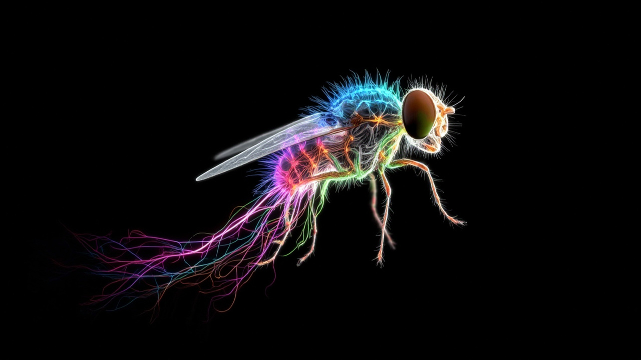
TL;DR: Scientists finished the first complete wiring diagram of an adult male fruit fly's brain and nerve cord, cataloging about 166,700 neurons and hundreds of millions of synapses.
Researchers announced on Sept. 3-4, 2026 that they had completed every neuron and connection in a male Drosophila fruit fly, giving neuroscience its second whole fly-brain map after a female map.
According to Ars Technica in "Second complete map of a fruit fly brain completed," the new male connectome joins a female Drosophila connectome finished previously, making the fly the first animal mapped twice at this resolution. According to the MRC Laboratory of Molecular Biology in "First complete connectome of male fly central nervous system allows for unprecedented male-female brain comparison," it is also the largest whole-brain connectome assembled so far.
The project was a years-long collaboration among the Howard Hughes Medical Institute's Janelia Research Campus, Google Research, and a Cambridge, U.K. team including the MRC Laboratory of Molecular Biology, the Cambridge Connectomics Group and the University of Cambridge Department of Zoology, with additional community collaborators.
Why a second fly map matters
A connectome is a parts list plus a wiring list: the position of each nerve cell in three dimensions and the synapses where cells exchange signals.
That wiring determines how sensation becomes action. Sensory cells detect light, sound, touch, smell and taste, interneurons transform and route those inputs, and motor cells drive movement, memory formation and other outputs. Without knowing which cell talks to which, researchers cannot trace how information travels.
The male map makes that tracing possible for both sexes. Because a female brain map already existed, the team could compare male and female circuits neuron by neuron and synapse by synapse across an entire adult nervous system.
The MRC Laboratory of Molecular Biology described the result as the first brain-wide comparison at synaptic resolution across sexes in an adult animal with complex anatomy and behavior. That comparison turns two reference maps into an experiment in itself: where the maps match, functions are likely shared; where they diverge, researchers have direct candidates for sex-specific behavior.
The new dataset is openly available as a public reference, so other laboratories can look up cell shapes, partners and pathways without rebuilding the map.
What's inside: 166,700 cells and millions of links
The male central nervous system map contains 166,700 neurons grouped into 11,710 identifiable cell types that recur across individual animals, according to the MRC Laboratory of Molecular Biology.
Google Research, a partner in the work, described the same scale in round terms as more than 166,000 neurons and nearly 12,000 distinct types. Ars Technica reported that the brain portion alone contains more than 300 million synaptic connections, even though the whole fly nervous system holds only roughly 150,000 to 166,000 cells in total.
The scale is difficult to grasp because each neuron branches. A single cell extends thin processes that can contact hundreds of partners. Counting cells is therefore only the start; charting every branch and contact is what creates hundreds of millions of entries.
Crucially, this connectome covers the brain together with the ventral nerve cord, the insect equivalent of a spinal cord, linked through an intact neck. The MRC Laboratory of Molecular Biology called that linkage a first for a fly and a critical advance because it permits end-to-end studies from sensory input in the head to leg and wing control in the body.
That continuity matters for behavior. Flies sense largely with head organs, including two large compound eyes and a retractable proboscis used for smell and liquid feeding. Signals from those sensors must travel through the brain and down the neck to motor neurons that steer walking, flight, feeding and courtship. A map that stops at the neck would leave that final leg invisible.
How thin slices became a 3D brain
The team started with a dissected central nervous system from one adult male fly and cut it into an extended series of extremely thin, evenly ordered sections.
Each slice was then imaged with high-resolution electron microscopy, producing millions of two-dimensional images detailed enough to show membranes and other fine cellular architecture. Because the slice order was known, software could stack the images back into a volume that preserved the three-dimensional organization of the brain.
Computers then carried the load that human tracers could not. Ars Technica noted that manual tracing with prints and markers, the approach used decades ago for a tiny roundworm, would be unworkable for a fly brain that is opaque and orders of magnitude more interconnected.
First, a generative model repaired the seams between slices. No blade passes without distortion and small material loss, leaving gaps that would confuse later steps. Google Research scientist Michal Januszewski told Ars Technica the model renders tissue as if those cuts were absent, allowing later processing to largely ignore the boundaries.
A second, recurrent model followed membranes through space to outline individual cells. Januszewski described it as moving through the volume while drawing neuron boundaries, a departure from familiar chat-style systems. Additional automated steps proposed synapses and cell classifications, followed by extensive human proofreading.
The MRC Laboratory of Molecular Biology estimated the combined automated and manual reconstruction effort as equivalent to 44 years of human labor. Artificial intelligence plus expert correction stitched millions of images into a coherent three-dimensional reconstruction of each neuron and its contacts.
Shared senses, different decisions
The headline biological finding is how little needs to change to produce different behavior.
Sensory and motor neurons are largely shared, or isomorphic, between males and females. Sex-specific cells found in only one sex and sexually dimorphic cells present in both but wired differently cluster instead in higher-order areas linked to decision-making and behavioral control, according to the MRC Laboratory of Molecular Biology.
There, they form dense, interconnected hubs that redirect otherwise common sensory streams into male or female behavioral circuits. The same smell, taste or visual cue can therefore trigger distinct responses.
Just under five percent of male brain neurons were classified as sex-specific or dimorphic, compared with fewer than three percent in females. Many of those cells express fruitless and doublesex, genes long tied to sex determination, shifting the emphasis from single influential cell types to system-wide circuitry.
Small numbers have outsized reach because those cells are highly connected. About 12 percent of neurons in the male brain show wiring differences as a result, against about 4 percent in the female brain, highlighting how a compact hub can reshape broader networks.
Google Research illustrated the idea with neuron type AOTU012, involved in sensory and taste processing. Paired AOTU012 cells exist in both sexes, yet connect to partly different neighbors in males and females, with some shared and some distinct partners.
From vision to courtship in one trace
The continuity through the neck lets researchers follow complete sensor-to-motor chains.
One example highlighted by Google Research runs from R1-R6 visual neurons in the eye through intermediate cells to DNg13, a motor neuron. The chain includes a male-specific interneuron called LoVP92, named for the so-called love spot region tied to courtship tracking.
That pathway connects environmental detection to movement in a single traceable route, showing how visual input can be routed toward sex-related pursuit behavior in males.
Three companion studies released alongside the map applied the resource to vision, taste and social behavior, according to Google Research. Rather than ending with publication, the connectome is intended as infrastructure for years of experiments on how circuits generate action.
From worm to fly to vertebrates
The project also refined methods the teams hope to extend toward larger nervous systems, potentially including vertebrates.
The historical contrast is stark. Work on the nematode C. elegans, with just over 300 neurons, began with hopes for machine vision in the early 1970s, then retreated to photographic prints and hand tracing when computers proved inadequate, Janelia senior group leader Gerry Rubin told Ars Technica. That worm map was eventually reported in 1986.
The fly effort reverses that history. The MRC Laboratory of Molecular Biology noted its lineage from the 302-neuron worm to a 3,000-neuron fly larva in 2023 and a whole adult female fly brain in 2024, now followed by the larger male brain-plus-cord map.
Each step demanded better sample preparation, imaging throughput, alignment software, segmentation models and proofreading workflows. The male project, built jointly by biologists who prepared and interpreted tissue and computer scientists who reconstructed hundreds of millions of contacts, tested that full pipeline at new scale.
Neither side could have finished alone, both teams acknowledged, according to Ars Technica. Biologists supplied intact anatomy at nanometer resolution; computing supplied the capacity to assemble millions of images into more than 166,000 consistent three-dimensional cells and their synapses in manageable time.
Sources referenced
- Second complete map of a fruit fly brain completed - Ars Technica (https://arstechnica.com/science/2026/09/brain-of-a-male-fly-completely-mapped-joins-earlier-map-of-a-female-brain/)
- First complete connectome of male fly central nervous system allows for unprecedented male-female brain comparison | MRC Laboratory of Molecular Biology (https://mrclmb.ac.uk/news-events/articles/first-complete-connectome-of-male-fly-central-nervous-system-allows-for-unprecedented-male-female-brain-comparison/)